Description
Interventional radiology hospital in Hyderabad
PACE Hospitals is One of the best Interventional Radiology Hospital in Hyderabad, offering effective minimally invasive treatment for disorders related to blood clots, blockages, tumours and chronic pain. The team of skilled and experienced interventional radiologists have vast expertise in managing a wide array of disorders requiring image-guided interventional treatment, including:
PACE Hospitals is one of the best hospitals for interventional radiology in Hyderabad. The department is staffed with a team of experienced interventional radiologists who coordinate closely with the multidisciplinary team of another department specialist to provide precise diagnostics and minimally invasive treatment for vascular disorders, oncological conditions, gastrointestinal conditions, urological issues and chronic pain management, to deliver the patient-centric and highest standard of care to the patients. Interventional radiology doctors are highly skilled in a comprehensive range of image-guided interventional and minimally invasive procedures including angioplasty, radiofrequency ablation, vascular stenting, embolization and more.
The Department of Interventional Radiology at PACE Hospitals is equipped with cutting-edge state-of-the-art diagnostic facilities, including CT scans, MRIs, and ultrasound-guided imaging, to evaluate and perform minimally invasive procedures to manage complex conditions. These procedures are intended to minimize hospital stay and discomfort and provide faster recovery.
What we treat?
We specialize in treating various critical conditions of blood clots, tumours, and blockages affecting the vascular system and other organs like the liver, kidney, lungs, uterus, digestive system, breast, prostate and bone & joints. From vascular conditions like Peripheral Artery Disease (PAD), Deep Vein Thrombosis (DVT), Varicose Veins, Aneurysms, Pulmonary Embolism to all kinds of cancer requiring radiofrequency ablation (RFA) and chemoembolization and conditions like Uterine Fibroids, Adenomyosis, Gastrointestinal Bleeding, Biliary Obstruction, Kidney Stones, Aortic Aneurysms, Coronary Artery Disease (CAD) requiring embolization and stent placement and solutions to the chronic pain management, Top Hospital for interventional radiology in Hyderabad is committed to delivering effective, precise and compassionate treatments with minimal recovery time.
Diagnostic Tests and Procedures of Interventional Radiology
We focus on providing precise interventional radiology diagnosis and treatment through minimally invasive techniques, offering patients an alternative to traditional surgeries. Our skilled and experienced interventional radiologist uses advanced, cutting-edge and high-precision imaging equipment like CT, MRI, and ultrasound to perform minimally invasive procedures, image-guided targeted treatment like angioplasty, ablations, tumour embolization, and catheter-based procedures for conditions including blood clots, tumours, and blockages without major surgery ensuring exceptional outcomes with quicker recovery and minimal discomfort.
1. CT scan (Computed Tomography): A CT scan utilizes an X-ray machine linked to a computer system to take a series of images of organs from different angles, which helps create detailed 3D pictures of the inside of the body.
2. MRI scan (Magnetic Resonance Imaging): An MRI is an imaging instrument that uses powerful radio waves and magnets to take images of the body in slices.
During the process, the patient has to lie on a table and is pushed into a long chamber, where the MRI makes loud thumping noises and rhythmic beats. A special dye (contrast agent) was injected into a vein, which shows the pictures more brightly.
3. Ultrasound: An ultrasound, also known as ultrasonography or sonography, utilizes high-energy sound waves to produce images of the internal organs of the body. When sound waves contact the body's organs, they are sent back by bouncing off them. A tool called a transducer turns the sound waves into images.
4. Angiography: Angiography is a medical imaging method that uses X-rays to check the blood vessels and heart chambers for anatomical and structural examination. A catheter is inserted into an artery through a small cut (incision) along with a contrast agent.
5. Angioplasty (Percutaneous transluminal coronary angioplasty) (PTCA): Angioplasty is a minimally invasive procedure to open (widen) the clogged narrowed coronary arteries to restore the regular blood flow to the heart muscle without opening the heart.
6. Stenting: Stenting is a minimally invasive technique made with fabric, metal, silicone mesh or materials, usually used to open and hold the open passages of the narrowed arteries. Stents treat narrowed coronary arteries (provides oxygenated blood to the heart muscle), aneurysm (bulge in the artery's wall) and narrowed airways in the lungs.
7. Embolization: Embolization is a minimally surgical treatment that helps close or block the blood vessels or abnormal blood channels. This is performed by introducing a substance called an embolic agent into the blood vessel to obstruct the flow of blood by using catheters.
8. Chemoembolization: Chemoembolization is a minimally surgical treatment for liver cancer that can be performed when the tumour cannot be treated by surgery or radio frequency ablation (RFA). During this process, chemotherapy drugs and embolic agents are injected into the tumour to block the arteries that supply blood (tumour).
9. Biopsy: In most cases, healthcare professionals must perform a biopsy to confirm the cancer. A biopsy is a minor procedure of removal of a sample of abnormal tissue, which is examined under the microscope and other examinations on the cell sample.
10. Fine-needle Aspiration: FNA (Fine-needle aspiration) is a procedure that healthcare professionals use to get a cell sample from an abnormal area or suspicious lump. It may also perform during a bronchoscopy or endoscopy.
It uses a thin needle and syringe to pull (take) out the abnormal cells, tissue and fluids. It will identify the masses (tumours) in the breast, skin, lymph nodes and thyroid.
11. Uterine Fibroid Embolization (UFE): Uterine Artery Embolization, or Uterine Fibroid Embolization (UFE), is a procedure to treat or reduce the size of symptomatic uterine fibroids (shrink non-cancerous tumours in the uterus) by blocking the blood supply.
12. Endometrial Ablation: Endometrial ablation is a minimally invasive procedure that destroys the endometrial tissue lining of the uterus to treat abnormal uterine bleeding. It can be caused by several reasons, such as hormonal changes, fibroids and polyps in the uterus.
13. Foreign Body Removal: If someone has an inhaled foreign object lodged in their airway, bronchoscopy is the preferred procedure to remove it. A healthcare professional may need to use rigid or flexible bronchoscopy to remove the object safely.
14. IVC filters: An inferior vena cava filter (IVC) is a tiny medical metal device placed in the inferior vena cava to prevent blood clots from travelling to the lungs. It is used to treat pulmonary embolisms. Healthcare professionals may recommend an IVC filter if the people have blood clots and cannot take blood thinners.
15. Cryotherapy: Cryotherapy is a medical procedure that utilizes extreme cold to freeze and eliminate abnormal cells. Healthcare professionals often use this technique to treat certain types of cancers, including lung cancer, by inducing tissue necrosis or cellular death.
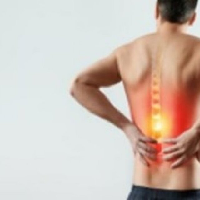
16. Vertebroplasty: When a vertebra fractures or breaks, it leads to the development of bone fragments. Pain will occur if they slide or rub against each other or push into the spinal cord. Vertebroplasty is a medical procedure that involves injecting the affected bone with a cement mixture, which is helpful to fuse the fragments or breaks, strengthen the vertebra and provide relief from the associated pain or discomfort.
17. Kyphoplasty: Vertebroplasty and kyphoplasty are medical procedures used to relieve the painful vertebral compression fractures in the spinal column, which can be caused by osteoporosis. A healthcare professional may use the imaging guidance for injecting the special cement mixture into fractured bone (known as vertebroplasty), or a balloon is inserted into the fractured or affected bone to create a space, which can then be filled with cement (known as kyphoplasty)
18. Nephrostomy: Kidneys produce urine, which flows through the ureter to the urinary bladder. Sometimes, urine flow is obstructed due to kidney stones, infections, cancers, trauma, etc. A nephrostomy is a medical procedure to clear out (drain) the urine from the kidney using a catheter (nephrostomy tube).
19. Cholangiography: Cholangiography is a procedure that involves the examination of the bile ducts and gallbladder structures by using a contrast dye to show them up on an X-ray. A healthcare professional sends the endoscope through the throat by numbing it.
20. Fluoroscopy: Fluoroscopy is an imaging method that visualises continuous X-ray images on the monitor screen. It studies moving body structures, much like an X-ray movie. Fluoroscopy can be used alone as a diagnostic tool or in combination with other procedures.
Why choose PACE Hospitals?
Expert Team
The interventional radiology department at PACE Hospitals consists of highly skilled radiologists, nurses, and support staff committed to providing personalized care. Our specialists work collaboratively with other departments, including oncology, cardiology, and gastroenterology, to ensure comprehensive treatment plans tailored to each patient's needs.
Commitment to Patient Care
At Multi specialty Hospital in Hyderabad, we prioritize patient comfort and satisfaction. From the initial consultation through post-procedure follow-up, our dedicated team is here to support patients every step of the way. We believe in clear communication and strive to provide patients and their families with all the information they need to make informed decisions about their healthcare.
Conclusion
Interventional radiology at PACE Hospitals, Hyderabad, represents a significant advancement in the treatment of various medical conditions. With a focus on minimally invasive techniques, advanced technology, and expert care, we are committed to improving patient outcomes and enhancing the overall healthcare experience.
Reviews
To write a review, you must login first.
Similar Items

Alaya Retreat Centre
The Injury and Performance Clinic
Full Body Checkup Price In Delhi| Qris Health
What antibiotic is good for dog skin infection?